Oral Cancer – Oral Screening
The stage that Oral Squamous Cell Carcinoma (OSCC) is diagnosed has a bearing on the outcome of OSCC; oro-pharyngeal cancers have relatively ‘silent’ symptoms which may not be present during the early stages of the disease, which is possibly why the stage of disease at diagnosis has not altered in the last 40 years despite public education.
For this reason, there is an interest in the early identification of OSCC yet the case for formal screening programmes does not meet the criteria established by the UK Screening Committee.
The Cochrane review on oral screening for OSCC found that overall there is not enough evidence to decide whether screening by visual inspection reduces the death rate for oral cancer and there is no evidence for other screening methods.
A GDP can expect to see at least a couple of mouth cancer lesions during their practising lifetime. Obviously, this is an average statistic affected by geographical location and social class of clientèle, much like the statistics for caries (tooth decay).
Nevertheless, to miss such a diagnosis could have a significant effect upon a patient’s health, quality and longevity of life and, of course, might leave the clinician open to criticism by the patient, and possibly give rise to medico-legal concerns.
One of the main problems with oral screening is limited accessibility to the dental office.
High risk groups are those less likely to have access to an NHS dentist and therefore less likely to have regular dental appointments.
These patients, many of whom are elderly, are less likely to schedule regular visits to the dentist due to financial constraints, a lack of adequate facilities or indifference towards their oral health.
Oral Cancer Screening
For oral cancer, where large numbers of patients are already seeing a dentist, an ‘opportunistic screening’ approach is generally advocated.
‘Opportunistic screening’ is less systematic but very much more cost-effective than population screening. If an opportunistic screening strategy is to be successful, all dentists should carry out the necessary soft tissue examination alongside hard tissue examinations.
Screening for oral cancer and pre-cancer becomes part of the routine examination. In practice, this will normally be at the beginning of each new course of treatment.
Cancers 2012 – Vision Paper, that by 2012, a pilot scheme should have been held in > 1 ‘spearhead PCT’ to
- setting up of a dedicated Head & Neck Clinic every 3 – 4 months to which over-50’s in high risk groups are invited
- links to specialists in secondary care, for example, sending digital photos for advice prior to a decision to refer although there are concerns about the feasibility of producing good enough quality photos
- posters in pharmacies offering mouth checks for ulcers
Also, action should have been taken with the manufacturers of products designed for self medication of oral ulcers and related conditions for the medication carton to carry a clear health warning about oral cancer. This will need to be managed carefully to avoid large numbers of the “worried well” overwhelming services.
Finally, the CRS opined that due to the problems of ‘dental capacity’ (which are outside the scope of the CRS to resolve), it may not be feasible to focus opportunistic screening with dentists. As a response to this perceived lack of capacity, it was thought that GP’s and practice nurses (who often see high risk groups for other issues such as high blood pressure) and pharmacists (who are often consulted about mouth ulcers) might be able to take up the slack.
Training issues and financial incentives would need to be considered as would the advantages / disadvantages of screening being carried out by non-medical personnel
Opportunistic Oral Cancer Screening
This was published by the British Dental Association in 2000.
This is quite comprehensive. It covers:
- Oral cancer screening – obligations and opportunities
- Risk factors
- Talking to patients about oral cancer
- Administration
- Examining the head, neck and soft tissue
- Using tolonium chloride
- Putting screening into practice
- If you were considering offering this type of screening service, it is well worth looking at.
It can be downloaded from here.
Oral Cancer Screening ‘Visual Aids’
To augment the GDP’s visual check of the mouth, there are a number of ‘aids’ to flag up suspicious areas.
These include:
- ViziLite & MicroLux DL
- Tolonium chloride (Toluidine Blue)
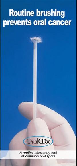
- OralCDx® brush biopsy
- Exfoliative cytology
Toluidine blue has been used for a number of years. ViziLite & OralCDx® are quite new. Exfoliative cytology is included for the sake of completeness but I don’t believe many GDP practices do this. This tends to be done in hospitals.
Most studies show that the various aids can help but when used for screening, economically, it is too expensive for the number of lesions correctly picked up.
VELscope, OralCDx® and Toluidine Blue staining have high false positive rates when they are used to screen routinely for oral cancer.
It would be inefficient to allocate scarce healthcare resources to the routine use of these devices for oral cancer screening.
These devices may be beneficial in opportunistic screening programmes or in cancer referral clinics when the pre-test probability of oral cancer is likely to be above 10%.
Further research is needed to determine at which pre-test probabilities, these adjunctive diagnostic devices would be cost-beneficial for the screening of oral cancer.
not a diagnostic tool.
In the UK, it is sold by Panadent. Checking their website, there seems to be a patchy take up over England; using this system, advertise their using this system, advertise their oral cancer screeningoral cancer screening facility.
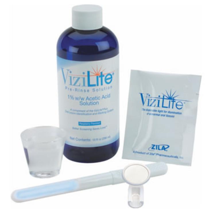
Kit contents:
• Chemi-luminescent device
• 30 ml acetic acid
• Light stick holder / retractor
Patient rinses with 1% acetic acid for 1 minute
Chemi-luminescent device activated & placed in ‘light stick’ holder.
Dim room lighting
Visually inspect oral cavity using device
Record any findings and refer the patient if necessary
How ViziLite® Works
Normal epithelium (skin) absorbs the light and appears dark; abnormal tissue reflects light and appears bright white. Based upon the current suggested usage for these devices, it is unclear what added benefit they would provide to the practicing clinician.
Toluidine Blue
- Meta-chromatic dye
- Stains nuclear DNA
- 1% aqueous solution followed by 1% acetic acid to de-colourise the oral lesion
- ‘Abnormal’ tissue retains the blue dye
Tolonium chloride or toluidine blue, is a stain that like Vizilite, is used as a screening device to help the clinician more easily visualise suspicious lesions.
Overall, toluidine blue appears to be good at detecting carcinomata but is positive in only ~50% of lesions with dysplasia. In addition, it also frequently stains common, benign conditions such as non-specific ulcers.
A systematic review concluded that there is no evidence that toluidine blue is effective as a screening test in a primary care setting.
The high rate of false positive stains and the low specific
All 3 layers (basal, intermediate and superficial layers) of epithelium are included / OralCDx® results come back as negative (no cellular abnormalities), positive (definitive cellular evidence of epithelial dysplasia or carcinoma) or atypical (abnormal epithelial changes warranting further investigation).
Negative lesions require the same careful clinical follow-up as negative histologically sampled lesions. Atypical and positive lesions require scalpel biopsy and histology analysis.
A report is sent with results for any specimens with atypical or positive findings. The report contains histological slides of the specimen and the oral pathologist’s report.
Disadvantages:
Often used inappropriately by the dentist. For example, some use it on papillomata.
Cells are seen out of context, which can result in misinterpretation. For example, if the Brush Test is used to collect cells from a patient who has lichen planus and the cells are spread out on a slide, they will be out of context and will be read as atypical.
Useful Articles & Websites
Homestead Schools, Inc (Dental)
International Agency for Research on Cancer / World Health Organisation
Oral Cancer Recognition Toolkit
BDA Occasional Paper 2000. Opportunistic Oral Cancer Screening.
BDJ 2003. Oral Cancer Prevention & Detection in Primary Healthcare.
Oral Oncology 2007. Review. Critical Evaluation of Diagnostic Aids for the Detection of Oral Cancer.
Am J Dent 2008. Review Article. Oral Cancer – Current & Future Diagnostic Techniques
JADA 2008. Oral Rinses may help detect HPV-positive Head & Neck Cancers
Evidence-Based Dentistry 2009. Editorial. Should we screen for Oral Cancer?
J Dent Res 2010. A community-based RCT for oral cancer screening with toluidine blue.
JADA 2010. For The Dental Patient…Detecting Oral Cancer Early
DHHS, NIH & NIDCR. Detecting Oral Cancer – A guide for health care professionals.ppt
Screening.nhs.uk 2010. Evaluation of Screening for Oral Cancer against NSC Criteria.
Evidence-Based Dentistry 2010. Clinical Recommendations for Oral Cancer Screening
Evidence Based Dentistry 2010. Editorial. Seek, don’t screen for Oral Cancer.
Evidence-Based Dentistry 2010. Does Toluidine Blue Detect More Oral Cancer
BDJ 2010. The Reality of Identifying Early Oral Cancer in the General Dental Practice
JADA 2010. Detecting Oral Cancer Early
Vital 2011. Making oral cancer screening a routine part of your patient care. Part 1
Vital 2011. Making oral cancer screening a routine part of your patient care. Part 2
BDA 2011. Early Detection of Oral Cancer. A Management Strategy
BJOMS 2011. Management of Oral Carcinoma. Benefits of early Pre-Cancerous Intervention
JADA 2011. Rise in Oral Cancer Linked to HPV, Study Shows
BDJ 2012. Microscope Created For Early Oral Cancer Diagnosis
BDJ 2016. Letters to the Editor. Mouth Cancer. Extending the RULE
BDJ 2018. Screening for Mouth Cancer – The Pros & Cons of a National Programme
BDJ 2018. Mouth Cancer – Presentation, Detection and Referral in Primary Dental Care


