Indices of Difficulty in Removing of 3rd Molars (Wisdom Teeth)
 Classification of impacted 3rd molars is often an attempt, based on 4 commonly used classifications of third molars, which are defined by angulation, impaction, application depth & eruption.
Assessment of difficulty of 3rd molar surgery is fundamental to forming an optimal treatment plan in order to minimise complications.
A compilation of both clinical and radiological information is necessary to make an intelligent estimate of the time required to remove a tooth and also whether the removal should be done in the dental surgery or a more specialised setting (such as a specialist clinic or hospital). Leading on from this is whether the tooth extraction would be better done under LA, LA Sedation or GA.
The various extraction difficulty indices include the following:
- Pell–Gregory classification
- Pederson scale
- Parant scale
- Winter’s Lines (WAR)
- WHARFE Scale
Various studies have shown that the Pell–Gregory scale, which is widely cited in textbooks of oral surgery, is not reliable for the prediction of operative difficulty.
Pederson proposed a modification of the Pell–Gregory scale that included a 3rd factor, the angulation of the molar (mesio-angular, horizontal, vertical or disto-angular). The Pederson scale is designed for evaluation of dental X-rays (such as DPT’s / OPG’s).
Although the Pederson scale can be used for predicting operative difficulty, it is not widely used because it does not take various relevant factors into account, such as bone density, flexibility of the cheek and buccal opening.
The Pederson scale is used in prediction of pre-operative difficulties. On the other hand, the modified Parant scale was implemented to predict post-operative difficulties.
Pre-operative Pederson scale (easy, moderate or difficult) and post-operative Parant scale (easy [I or II] or difficult [III or IV]).
Winter’s Lines (WAR)
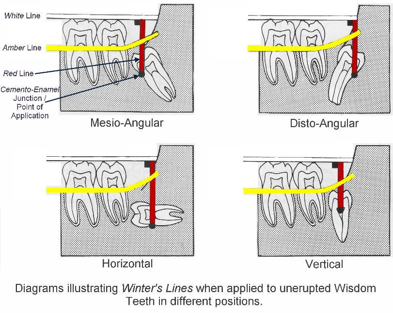
The position & depth of the mandibular 3rd molar can be determined using the Winter’s Lines (WAR). These are 3 imaginary lines (red, amber & white) “drawn” on the dental X-ray (these days, normally an OPG / DPT).
White Line
The white line is drawn along the occlusal surfaces of the erupted mandibular molars & extended over the 3rd molar posteriorly. It indicates the difference in occlusal level of the 1st & 2nd molars & the 3rd molar.
Amber Line
The amber line represents the (height of the) bone level. The amber line is drawn from the surface of the bone on the distal aspect of the 3rd molar (or from the ascending ramus) to the crest of the inter-dental septum twixt the 1st & 2nd molars. This line denotes the margin of the alveolar bone covering the 3rd molar and gives some indication to the amount of bone that will need to be removed for the tooth to come out.
Red Line
The red line is an imaginary line drawn perpendicular from the amber line to an imaginary point of application of an elevator. Usually, this is the cemento-enamel junction on the mesial aspect of the impacted tooth (unless, it is the disto-angular impacted tooth where the application point is the distal cemento-enamel junction). The red line indicates the amount of bone that will have to be removed before elevation of the tooth i.e. the depth of the tooth in the jaw & the difficulty encountered in removing the tooth.
With each increase in length of the red line by 1mm, the impacted tooth becomes 3 x more difficult to remove (as opined by Howe). If the red line is < 5mm, than the tooth can be removed under just LA; anything above, a GA or LA Sedation would be more appropriate.

 Another method of judging the depth of the 3rd molar is to divide the root of the 2nd molar into thirds. A horizontal line is drawn from the point of application for an elevator to the 2nd molar. If the point of application is adjacent to the coronal, middle or apical root third, then the tooth extraction is assessed as easy, moderate or difficult respectively.
WHARFE Assessment
The six factors chosen for scoring are:
- Winters classification
- Height of the mandible
- Angulation of the 2nd molar
- Root shape & morphology
- Follicle development
- Path of Exit of the tooth during removal
The scoring by this system helps the beginners to anticipate problems and to avoid difficult impactions. Unfortunately, the disadvantage of this method is that it is related only to radiological features alone; the details of the surgical procedures are not considered. The total scoring is directly related corresponding difficulties in removing that impacted teeth.
Assessment of difficulty of third molar surgery is fundamental to forming an optimal treatment plan in order to minimise time required to remove a tooth and whether it would be better done just under LA or under LA Sedation or GA.
There are a number of classifications / scales that try to be predictive of the extraction however each has its good and bad points.
There has been an attempt to computerise the assessment of impacted 3rd molars. However good this is though, there is still the problem of whether the scale used is of any use or widely understood.
The acid test for any of these classifications / scales is whether they are actually used in OMFS Departments or dental surgeries. From personal experience, they are not.
Where the various classifications are not used, the following observations are more likely to be noted and acted upon.
Factors that Make Surgery Less Difficult
- Mesio-angular impaction
- Class 1 ramus
- Class A depth
- Roots 1/3 – 2/3 formed (present in the younger patient)
- Fused conical roots
- Wide periodontal ligament (present in the younger patient)
- Large follicle (present in the younger patient)
- Elastic bone (present in the younger patient)
- Separated from 2nd molar
- Separated from IDN
Factors that Make Surgery More Difficult
- Disto-angular impaction
- Class 3 ramus
- Class C depth
- Long thin roots (present in the older patient)
- Divergent curved roots
- Narrow periodontal ligament (present in the older patient)
- Thin follicle (present in the older patient)
- Dense, inelastic bone (present in the older patient)
- Contact with 2nd molar
- Close to IDN
- Complete bony impaction
Useful Articles & Websites
BDJ 2001. Factors predictive of difficulty of mandibular third molar surgery
BJOMS 2002. Classification of Surgical Difficulty in Extracting Impacted 3rd Molars
JOMS 2004. Risk Factors for 3rd Molar Extraction Difficulty
JOMS 2005. How Well Do Clinicians Estimate 3rd Molar Extraction Difficulty?
BJOMS 2007. Pederson scale fails to predict how difficult it will be to extract lower 3rd molars


